Approaches
Magnetic Resonance Imaging and Transcranial Magnetic Stimulation (TMS) have both immense potential in the field of neuroscience:
- Structural and functional MRI allow for mapping cognitive functions to spatial and temporal features of brain activation.
- Transcranial Magnetic Stimulation holds the potential to infer on causal relationships between brain areas and cognitive functions.
- Transcranial Magnetic Stimulation and fMRI can support each other based on their respective advantages when combined.
fMRI before, after and during brain stimulation
Magnetic resonance imaging complements brainstimulation in different ways
- Anatomical images acquired before stimulation can be fed into the neuronavigation software to define the actual stimulation site (for neuronavigated TMS).
- neuroimaging before stimulation allows for functional definition of a stimulation target.
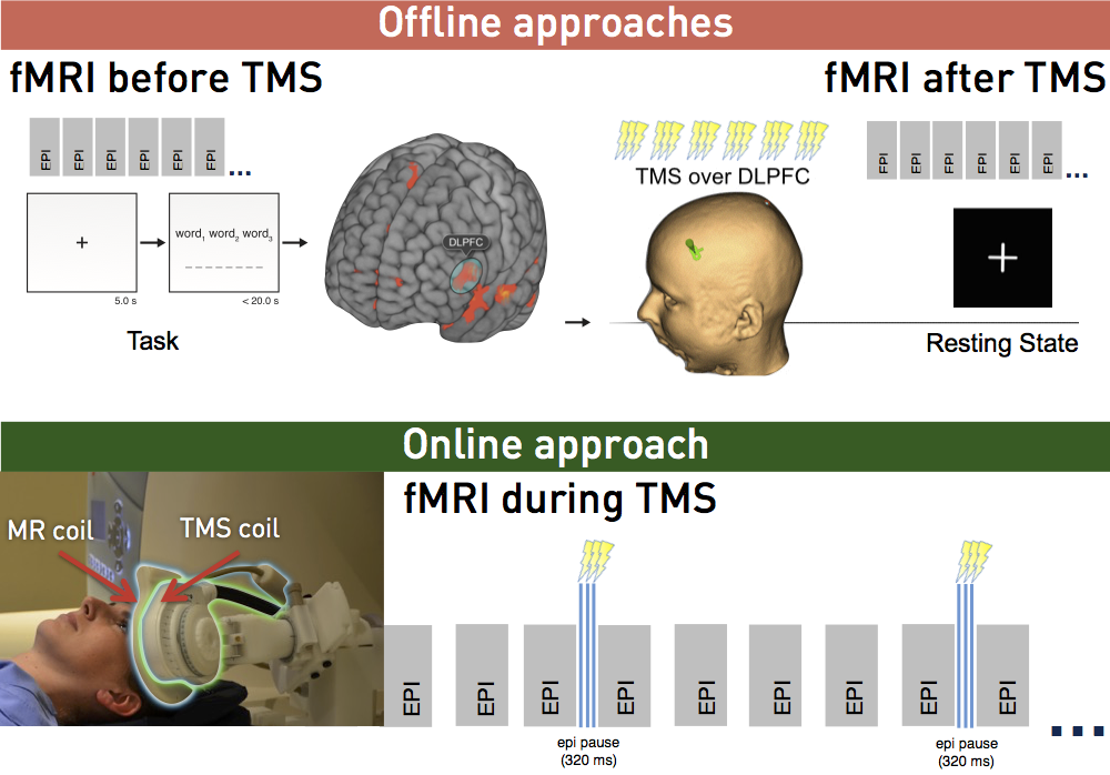
- Offline imaging (e.g. fMRI during task or in resting-state) allows for evaluating long-term effects after brain stimulation.
- Finally, fMRI during stimulation, i.e. interleaved TMS-fMRI, allows for the direct observation of TMS-induced effects.
Concurrent TMS/fMRI
Methods for concurrent use of imaging and TMS were developed to examine evoked responses in detail. This combination allows for the investigation of direct and localized neural effects as well as network effects due to stimulation, yielding studies that go beyond simple observation of behavioural changes. There are, however, a number of challenges that have to be addressed for successful imaging of online effects.
Why interleaved TMS-fMRI?
One of the primary advantages of interleaved TMS-fMRI is its ability to investigate causal relationships between brain regions. While fMRI alone can show correlations between brain activity and behavior, it cannot establish causality. Brain stimulation techniques, on the other hand, can directly influence neural circuits, helping researchers pinpoint the effects of stimulating specific areas on overall brain function. This is particularly valuable in studying brain disorders, such as depression, epilepsy, and stroke, where abnormal connectivity or dysfunction in certain brain areas can be better understood.
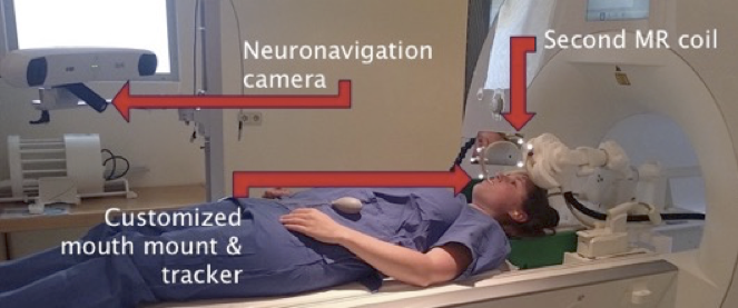
Our concurrent imaging & stimulation setup has a high-density of multi-channel receive coils that are positioned directly on the subject’s scalp to maximise signal to noise ratio. This is achieved by either surface coils sandwiched between head and stimulation coil or a ultra-thin flexible coil that can be wrapped around the head before magnetic stimulation pulses are applied through it.
read more
We have developed and patented a dedicated concurrent TMS/fMRI coil array.