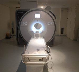
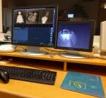
 Functional Magnetic Resonance Imaging (fMRI) is a neuroimaging technique that allows for non-invasive investigation of brain function. Functional MRI uses MR imaging to measure the tiny metabolic changes that take place in more active parts of the brain: Using radiofrequency pulses and a strong magnetic field, images of the brain can be acquired. Functional MRI has become the method of choice for various neuroscientific and clinical research questions as well as routine applications.
Mapping Brain Function
When you engage in simple motor tasks (e.g. finger movement), visually perceive certain stimuli (see retinotopic mapping) or when you have a creative insight (Aha-moment), certain regions of your brain show increased activation. This leads to a higher energy-demand and therefore blood oxygenation, which in turn allows for mapping those functions using neuroimaging.
Brain activation changes during finger-movement. (see Windischberger et al., 2008)
In a classic task-based fMRI experiments, participants are lying inside an MR scanner and images are acquired continuously (neuroimaging) while the participant is engaged in a task. Via measuring changes in blood oxygenation level dependent (BOLD) MR signal and modeling the influence of specific task conditions (e.g. periods in which subjects are moving their finger compared to rest), brain regions that are associated with these specific functions can be identified.
Imaging Brain Network Connectivity

Functional connectivity map (default mode network)
The brain is still highly active when you close your eyes and think of nothing special. In this so-called resting state certain brain regions are synchronised, i.e. they show a highly correlated fluctuation in activation level. Since regions that are active together are likely connected to each other, this allows for revealing brain network connectivity properties. The default mode network (see figure right) is one of the most commonly studied networks. Hot colors indicate regions that show a high level of synchrony and are therefore very likely functionally connected.
Clinical Neuroimaging
Besides mapping brain function and identifying networks associated with specific cognitive processes in healthy volunteers, patients can be examined to identify brain regions that are associated with functions impaired by neuropsychiatric diseases. Given the possibility of high spatial resolution in the range of about 1 to 5 mm³ and its capability to measure cortical and sub-cortical structures, fMRI at ultra-high field has become a gold standard neuroimaging method for investigating neural mechanisms. Thus, fMRI neuroimaging can be considered an indispensable tool for both fundamental basic and applied research in cognitive science, neuroscience, psychology, and other life science disciplines.
fMRI Routine Applications
 Functional MRI has evolved into an invaluable clinical neuroimaging tool for routine brain imaging, fostered by continuous improvement of preclinical research and technical progress (bench-to-bedside). Its applications have advanced the clinical standard of diagnosis, pre-surgical localization of brain functions, and progress monitoring.
Multimodal Neuroimaging & Limitations
The integration of fMRI and other neuroimaging techniques, e.g. Positron Emission Tomography (PET) is promising since it allows to e.g. link changes in fMRI brain activation (BOLD) to specific neurotransmitter binding. However, all pure imaging approaches are limited to correlational research questions. It is unclear if a brain area showing increased activation during a task period is causally involved in a cognitive task or rather a correlate of a specific affective component. To investigate and establish causal relationships between the modulation of neuronal activity and following changes in cerebral function and overt behavior, other methods such as non-invasive brain stimulation / neurostimulation are needed.
Neuroimaging & Neurostimulation
Neurostimulation methods such as transcranial magnetic stimulation (TMS) have the potential to introduce causality into imaging studies, since they allow to transiently manipulate activity in specific brain areas. However in the current state-of-the-art practice the response rate for brain stimulation is limited. A better understanding of the mechanism of actions of TMS will help to improve its application.
The combination of TMS brain stimulation with resting-state fMRI has been shown to promote the advances in both fields: in terms of connectivity research TMS can help to understand the interactions between remote brain areas on a new causal level, while network measures can promote guiding and evaluation of TMS application and improve stimulation protocols.
Read more about Combining TMS with fMRI
Concurrent TMS/fMRI
Ultimately the realtime measurement of TMS evoked brain activation changes (BOLD) is possible using a novel dedicated concurrent TMS setup developed by our group. Its a highly promising tool with the potential to e.g. adjust stimulation parameters (e.g. dose) for the individual subject / patient to improve response rates in science and clinical precision-medicine.

